Vascular Access
IN THIS LESSON
Central Venous Access - Lesson Overview
What Is Central Venous Access?
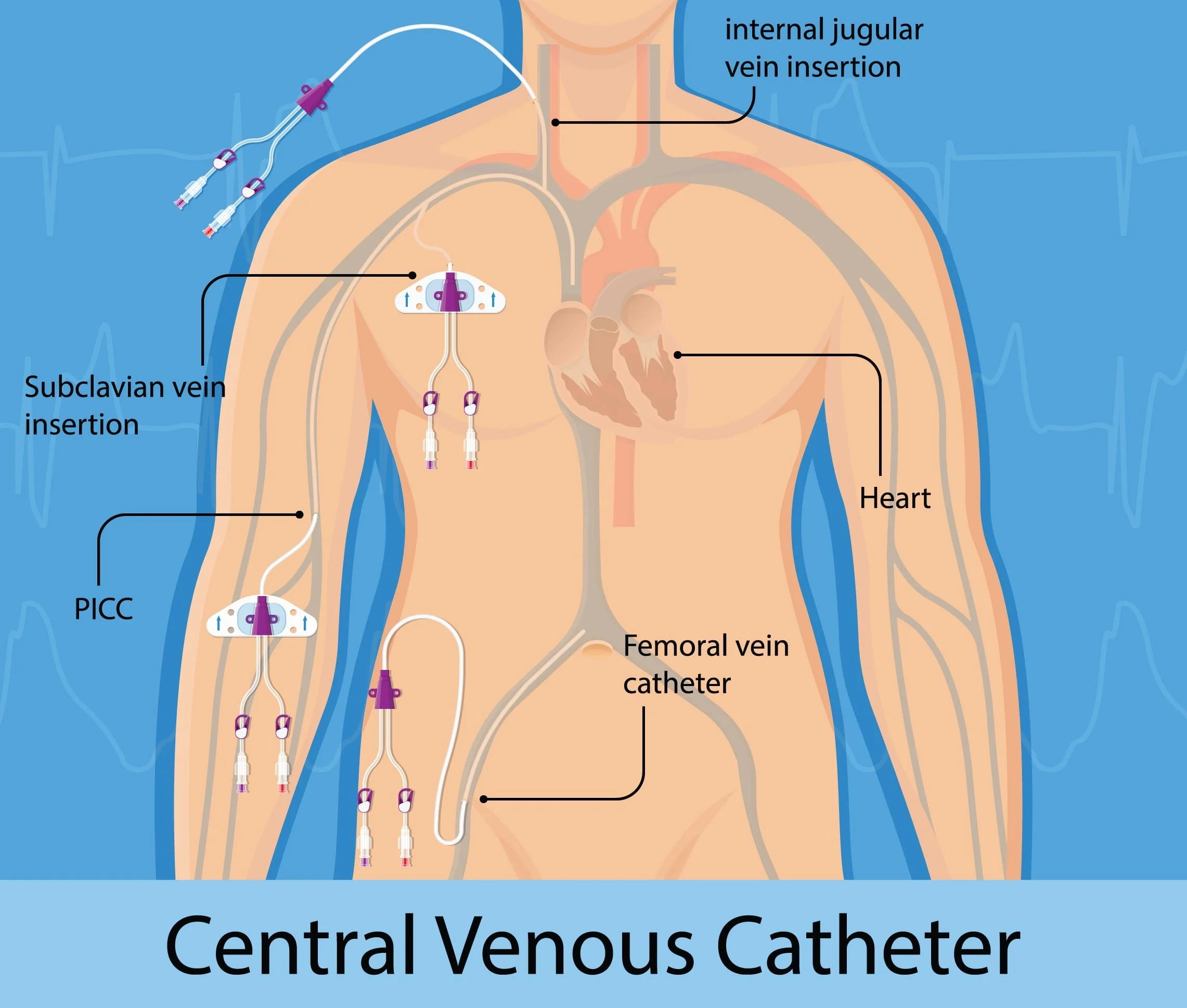
Central venous access refers to the placement of a catheter into a large, central vein — most commonly the internal jugular, subclavian, or femoral vein — so that its tip rests near the right atrium in the superior or inferior vena cava. Unlike peripheral IVs, central venous catheters (CVCs) provide direct access to high-flow vessels close to the heart, making them indispensable in both acute and long-term patient care.
CVCs are placed for a wide range of clinical indications, including administration of vasopressors, total parenteral nutrition (TPN), chemotherapy, and long-term antibiotics, as well as for hemodynamic monitoring and emergent resuscitation. In interventional radiology, these procedures are performed using real-time imaging guidance — typically ultrasound and fluoroscopy — which dramatically improves safety and success rates compared to landmark-based techniques.
What You'll Learn in This Video
By the end of this lesson, you will be able to:
Identify the indications and contraindications for central venous catheter placement, including when IR-guided access is preferred over bedside insertion
Recognize the relevant anatomy of the major central venous access sites — internal jugular, subclavian, axillary, and femoral veins — and understand why site selection matters
Understand the equipment used in IR-guided central line placement, including catheter types (standard CVC, PICC, tunneled catheter, port-a-cath) and their clinical applications
Walk through the procedural steps of ultrasound-guided venipuncture and the Seldinger technique, from skin prep to catheter securement
Recognize intra-procedural imaging landmarks used under fluoroscopy to confirm appropriate catheter tip position
Identify common complications such as pneumothorax, arterial puncture, air embolism, and catheter malposition — and understand how they are recognized and managed
-
Add a short summary or a list of helpful resources here.